Heart Rate Changes During Brain Stimulation Predict Depression Treatment Success Weeks Later

Depression and cardiac biomarkers in brain stimulation therapy. (A) Depression affects millions worldwide, with at least one-third of patients not responding well to conventional treatments. (B) Electrocardiogram patterns during brain stimulation.
Cardiac monitoring during first session forecasts six-week outcomes in major depression, challenging personalized targeting approaches.
Researchers at the University Medical Center Göttingen tracked beat-to-beat heart rate changes in 75 patients with major depressive disorder during accelerated intermittent theta burst stimulation (iTBS). Patients whose heart rates slowed more dramatically during initial treatment showed significantly greater reductions in depressive symptoms six weeks later, with the correlation holding specifically for active stimulation but not sham treatment.
"Patients who showed greater heart rate deceleration within the first 45 seconds of initial stimulation demonstrated superior clinical improvement at the six-week follow-up," the research team led by Dr. Roberto Goya-Maldonado reported. The relationship achieved statistical significance (r=0.27, P=0.021) and was significantly stronger than the sham condition, suggesting the cardiac response reflects meaningful engagement of mood-regulating brain circuits.
The findings arrive as clinicians worldwide seek better methods to predict and optimize outcomes for treatment-resistant depression, which affects approximately one-third of the 16-20% of the population diagnosed with major depressive disorder during their lifetimes.
Challenging Personalized Targeting Assumptions. The study simultaneously tested a widely promoted approach: personalizing treatment location based on individual brain connectivity patterns. Surprisingly, this sophisticated neuroimaging method showed no advantage over simple standardized positioning.
Researchers randomized patients to receive stimulation either at personalized sites identified through resting-state functional MRI scans or at the standard F3 location determined using the international 10-20 EEG system. Despite advanced MRI technology mapping each patient's unique brain connectivity between the left dorsolateral prefrontal cortex and the default mode network, personalized targeting produced equivalent symptom reduction.
The distance between stimulated sites and theoretically optimal targets averaged 7.27±4.76 millimeters for personalized targeting compared to 18.20±9.17 millimeters for F3 positioning. Yet this twofold difference in spatial precision yielded no measurable clinical benefit. Correlations between distance to ideal sites and clinical improvement remained non-significant for both active (r=0.17, P=0.14) and sham (r=0.08, P=0.48) conditions.
"While personalized targeting based on connectivity may be disappointing, cardiac biomarkers offer a new and practical path toward treatment optimization," wrote Dr. Julio Licinio and Dr. Helen Mayberg in an accompanying editorial.
How Heart Rhythms Reflect Brain Circuit Engagement. The cardiac deceleration likely reflects successful activation of the frontal-vagal pathway, a neural circuit connecting the prefrontal cortex to the heart through the subgenual anterior cingulate cortex and brainstem. When brain stimulation effectively engages mood-regulatory networks, signals propagate through subcortical structures to brainstem vagal nuclei, triggering measurable heart rhythm changes.
Previous research in healthy volunteers demonstrated that F3 stimulation optimally induces such heart rate changes, with preliminary depression studies showing trends toward associations between cardiac modulation and clinical response. The current trial provides the first definitive statistical evidence linking early cardiac changes to long-term symptom improvement.
However, the relationship proved complex. While heart rate deceleration (RR interval slope) predicted six-week outcomes, changes in heart rate variability during stimulation showed unexpected inverse relationships with short-term improvement.
The root mean square of successive differences between heartbeats (RMSSD)—a standard heart rate variability measure—increased during active compared to sham stimulation as anticipated. But patients showing smaller RMSSD increases during the first 270 seconds experienced greater symptom reduction at one-week assessment (r=-0.29, P=0.013).
"That was unexpected," the researchers acknowledged. They proposed that effective frontal-vagal engagement may initially reduce variability during stimulation, followed by compensatory increases aligning with clinical improvement. However, this explanation remains speculative, highlighting gaps in understanding brain-heart interaction dynamics during neuromodulation.
Linear mixed model analysis revealed that the interaction between active stimulation and RMSSD change significantly predicted within-week symptom improvement at 180 seconds (β=-0.015, P=0.011) and 270 seconds (β=-0.017, P=0.008). The negative coefficients indicate that greater stimulation-specific increases in heart rate variability corresponded with worse short-term outcomes.
Study Methodology and Safety Profile: The quadruple-blind, sham-controlled crossover trial enrolled 92 patients with major depressive disorder between April 2019 and July 2021, with 75 completing the full protocol. Participants received either personalized or F3 stimulation positioning, in active-sham or sham-active sequences.
The accelerated iTBS protocol delivered intense treatment over two one-week periods. Each daily session consisted of four stimulation blocks, with patients receiving 7,200 pulses daily and 36,000 pulses across the treatment week. Stimulation parameters followed established protocols: two seconds on, eight seconds off, with volleys of 10 bursts at 5 Hz intensity set at 110% of individual resting motor threshold.
Continuous electrocardiogram monitoring throughout sessions captured beat-to-beat heart rate changes. The research team used three chest electrodes recording at 1,000 Hz with precise synchronization to iTBS bursts. Data preprocessing involved removing noise, detecting R-peaks, calculating RR intervals, and correcting for ectopic beats using validated algorithms.
Sham conditions employed sophisticated blinding. The magnetic coil rotated 180 degrees according to pre-coded sequences unknown to patients, providers, or raters. Simultaneously, transcutaneous electrical nerve stimulation electrodes on the scalp mimicked tingling sensations timed with stimulation sounds. Post-treatment assessments revealed no significant expectation differences between active (0.43±0.29) and sham (0.38±0.29) conditions (P=0.15), confirming successful blinding.
Safety monitoring documented headache (53.33%), neck pain (30.00%), scalp pain (58.00%), and scalp irritation (20.67%). However, average daily intensity remained very low on 0-10 scales in both active and sham conditions. Only headache showed statistically significant differences between conditions (P=0.007). No participants reported serious adverse effects.
The authors provided some clinical context and treatment landscape. Major depressive disorder represents one of the leading causes of disability worldwide, affecting 16-20% of the population over their lifetimes. Conventional antidepressant medications fail to produce adequate response in approximately one-third of patients, even after multiple trials. For these treatment-resistant cases, brain stimulation therapies offer important alternatives with highly variable outcomes.
Repetitive transcranial magnetic stimulation uses powerful magnetic fields to induce electrical currents in specific brain regions without surgery. Recent advances in stimulation protocols, particularly intermittent theta burst stimulation with accelerated scheduling, have expanded clinical adoption.
Response rates to standard protocols currently range from 30-50%. This variability has driven intense biomarker research. Various approaches have been proposed, including neuroimaging-based targeting, electroencephalography patterns, genetic markers, and symptom profiles. Few have demonstrated sufficient predictive accuracy or practical feasibility for routine implementation.
The cardiac monitoring approach, termed neuro-cardiac-guided transcranial magnetic stimulation, proposes that left dorsolateral prefrontal cortex stimulation triggers measurable autonomic effects through pathways to brainstem vagal nuclei. Compared to healthy controls, depressed patients exhibit altered heart rate and reduced heart rate variability, though these changes do not always normalize with treatment.
Why did standard measurements correlated differently? The selective association of cardiac biomarkers with Montgomery-Åsberg Depression Rating Scale scores—but not Hamilton Depression Rating Scale or Beck Depression Inventory scores—illuminates another complexity. Different assessment instruments emphasize distinct symptom domains.
Factor analyses show the Montgomery-Åsberg scale loads heavily on observed mood symptoms, the Hamilton scale on neurovegetative features like sleep and appetite, and the Beck Inventory on cognitive symptoms. That cardiac biomarkers specifically predict mood improvements, but not other symptom clusters, suggests the heart rate response reflects particular underlying mechanisms potentially enabling more targeted treatment selection.
What are the implementation challenges and practical barriers? Despite promising findings, several obstacles complicate connectivity-based targeting translation. The personalization approach requires expensive MRI scanning, specialized analysis expertise, sophisticated neuronavigation systems, and time-intensive planning—costs and demands substantially exceeding standard positioning methods.
Moreover, achieving precise targeting proved difficult even with advanced technology. Actual stimulation sites in the personalized group deviated from calculated targets by more than 10 millimeters in some participants, reflecting real-world challenges including coil positioning constraints, anatomical variations, and patient tolerance. Such deviations could dilute potential benefits, though the lack of correlation between accuracy and outcomes suggests spatial precision may matter less than believed.
In contrast, cardiac monitoring requires only electrocardiogram equipment already standard in medical settings, involves minimal additional time, and provides immediate feedback. If clinicians could adjust stimulation parameters based on real-time cardiac responses within the first session, treatment optimization might occur immediately rather than after weeks of assessment.
"Equipment costs, training requirements, and workflow integration pose practical barriers even if the science proves robust," the editorial authors cautioned. The field continues searching for psychiatry's equivalent of HER2 testing in breast cancer—biomarkers that fundamentally alter treatment decisions and improve outcomes.
Study Limitations and Unanswered Questions. The research team transparently acknowledged several limitations. Depression's heterogeneity likely obscures group-level effects. Different symptom profiles may respond to specific stimulation targets. With 75 participants, the study lacked power to identify such subgroups or determine whether personalized targeting benefits particular patient populations.
The crossover design strengthened internal validity but complicated long-term interpretation. The six-week assessment corresponded to approximately five weeks after active stimulation for one group but only three weeks for the other. Future parallel-group studies with consistent follow-up could clarify whether personalized targeting benefits emerge at specific windows.
Cardiac measurement approaches also require standardization. The study assessed heart rate variability during stimulation rather than at rest, capturing stimulation-induced changes rather than baseline tone. No consensus exists regarding optimal cardiac assessment protocols during neuromodulation.
The researchers could not replicate findings with secondary outcome measures. While they documented relationships between cardiac parameters and Montgomery-Åsberg scores, the lack of associations with other depression scales suggests these biomarkers may be more limited or symptom-specific than hoped.
Critical questions remain: Will combining cardiac monitoring with other biomarker approaches improve prediction accuracy? Can real-time cardiac feedback enable active parameter adjustments during sessions, creating closed-loop optimization? How do individual differences in autonomic function, cardiovascular health, or medications influence these relationships?
Implications for Depression Neurobiology. The contrasting results for neuroimaging-guided targeting versus cardiac biomarkers raise fundamental questions about depression pathophysiology and therapeutic target engagement measurement. If cardiac responses predict improvement better than brain connectivity measures, peripheral physiological markers may capture integrative processes that focal brain imaging misses.
Alternatively, the relative simplicity of electrocardiogram signals compared to functional MRI may produce less noisy measurements rather than reflecting fundamentally different biology. Resting-state connectivity shows considerable within-individual variability, potentially limiting treatment targeting utility.
Another possibility is that the prefrontal-subgenual cingulate connectivity model represents an incomplete picture. More comprehensive approaches incorporating autonomic pathways, multiple brain networks, or different connectivity measures might yield stronger targeting signals.
The unexpected inverse relationship between heart rate variability changes and short-term improvement particularly challenges prevailing models. Lower heart rate variability represents a well-established pathological finding in depression, generally indicating reduced parasympathetic activity. Conventional wisdom suggests increasing heart rate variability should align with improvement. The observation that smaller increases during stimulation correlated with better outcomes contradicts this assumption.
What are the future research priorities? Replication in larger, more diverse samples will establish whether results generalize across populations, settings, and regions. The current study enrolled primarily European patients at a single academic center; broader validation should include varied racial and ethnic backgrounds, socioeconomic circumstances, depression severity levels, and comorbidity profiles.
Head-to-head comparisons of different biomarker approaches—cardiac, electroencephalographic, neuroimaging, genetic, inflammatory—could identify optimal prediction strategies or reveal complementary combinations. Some biomarkers might predict overall response while others guide parameter selection.
Mechanistic studies using concurrent neurophysiological recordings might clarify how cardiac responses relate to neural circuit dynamics. Simultaneous electroencephalography, functional near-infrared spectroscopy, or functional MRI during stimulation could map pathways from cortical activation through subcortical structures to brainstem centers and peripheral cardiac effects.
Most critically, clinical trials directly comparing biomarker-guided protocols with standard protocols will determine whether physiological monitoring improves patient outcomes. Do algorithms adjusting coil positioning, intensity, or frequency based on cardiac feedback produce higher response rates or faster improvement? Only randomized comparisons with clinical endpoints can establish whether added complexity justifies implementation.
The field must address practical questions: What cardiac response threshold should trigger adjustments? How should clinicians modify treatment when initial signals appear suboptimal? Should different patient subgroups receive different monitoring approaches? Can simplified systems work in typical settings without research-grade equipment?
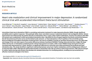
About the Research and Editorial: The research article, "Heart rate modulation and clinical improvement in major depression: A randomized clinical trial with accelerated intermittent theta burst stimulation," was authored by Jonas Wilkening, Henrike M. Jungeblut, Ivana Adamovic, Vladimir Belov, Peter Dechent, Lara Eicke, Niels Hansen, Vladislav Kozyrev, Lara E. Marten, Yonca Muschke, Caspar Riemer, Knut Schnell, Asude Tura, Melanie Wilke, Fabian Witteler, Jens Wiltfang, Anna Wunderlich, Valerie Zimmeck, Anna Zobott, Carsten Schmidt-Samoa, Tracy Erwin-Grabner, and Roberto Goya-Maldonado from the University Medical Center Göttingen and affiliated institutions.
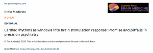
The accompanying editorial, "Cardiac rhythms as windows into brain stimulation response: Promise and pitfalls in precision psychiatry," was written by Dr. Julio Licinio, Editor-in-Chief of Genomic Press, and Dr. Helen S. Mayberg of the Icahn School of Medicine at Mount Sinai.
Both articles are freely available via open access in Brain Medicine at https://doi.org/10.61373/bm025a.0113 (research article) and https://doi.org/10.61373/bm025d.0119 (editorial).
The study was supported by the German Federal Ministry of Education and Research (BMBF: 01 ZX 1507, "PreNeSt - e:Med"). The study protocol was approved by the Ethics Committee of the University Medical Center Göttingen and registered at clinicaltrials.gov (NCT05260086). The authors declared no conflicts of interest.
For further research information - contact: Laboratory of Systems Neuroscience and Imaging in Psychiatry (SNIP-Lab) Department of Psychiatry and Psychotherapy University Medical Center Göttingen Von-Siebold-Straße 5, 37075 Göttingen, Germany Email: roberto.goya@med.uni-goettingen.de
Ma-Li Wong
Genomic Psychiatry
maliwong7@gmail.com
Legal Disclaimer:
EIN Presswire provides this news content "as is" without warranty of any kind. We do not accept any responsibility or liability for the accuracy, content, images, videos, licenses, completeness, legality, or reliability of the information contained in this article. If you have any complaints or copyright issues related to this article, kindly contact the author above.


